animal cell under microscope labeled
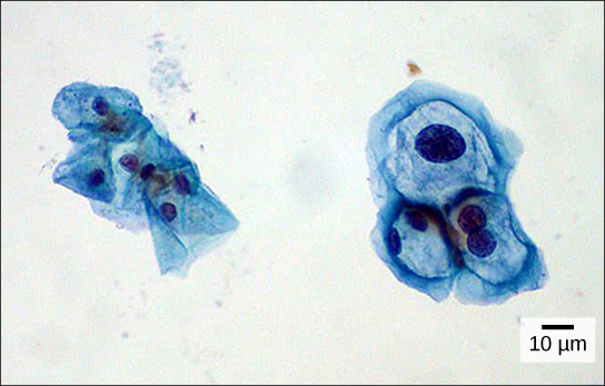
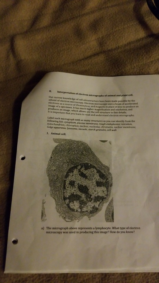
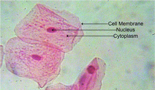
In this book he gave 60 observations in. The large spherical area is the nucleus while the granulated part is the cytoplasm of the cell.
Typical Animal Cell And Plant Cell Hi Res Stock Photography And Images Alamy
As per the given information in the question cells of mushrooms plants and animals all have visible nuclei under a microscope.
. Add a drop of purple stain specific for animals and cover with a cover slip. Lab Using A Microscope. Hair under microscope.
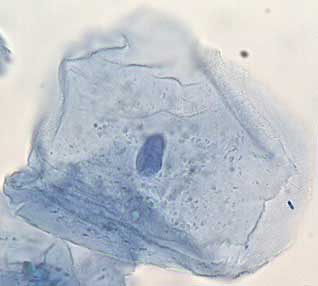
Draw a diagram of one cheek cell and label the parts. Here the seminiferous tubules of the animal show different types of cells like primary spermatocytes secondary spermatocytes spermatid and spermatozoa. Neuron under microscope labelled diagram.
Simple columnar epithelium under a microscope The simple columnar epithelium under a microscope consists of a single layer of taller than wide cells. Students will observe onion cells under a microscope. So hair is an epidermal down growth embedded into the dermis or hypodermis of the animals skin.
You will find two main parts in hair a cylindrical. The first animal cell was observed under an optical microscope which clearly showed the nucleus and microfilament network in. Animal Cell Under A Microscope Labeled - What is a diagram of a plant and animal cell under an.
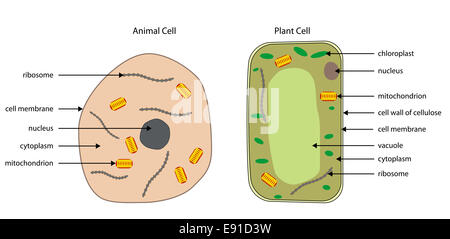
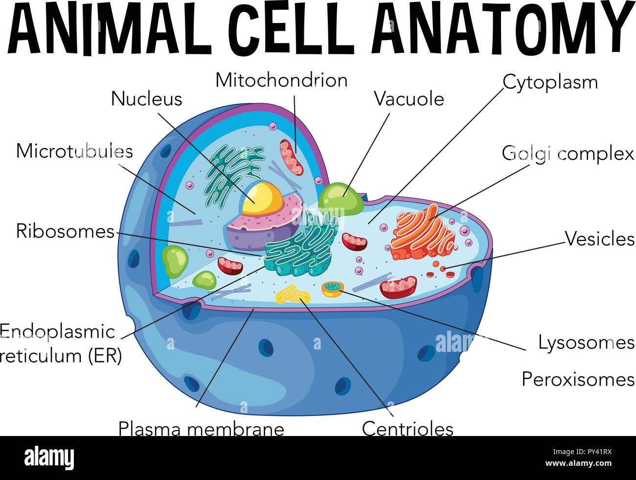
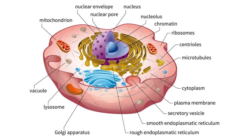
Cell plant structure animal microscope diagram generalized under electron cells labeled basic class biology function plants notes diagrams living light. Elodea Plant Cell Under Microscope Labeled. The cell membrane the nucleus and the cytoplasm.
- Jun 10 2021 based on my minimal understanding viruses are too. Beginning with a cell spread on the substrate follow prophase. Gently roll and rub the toothpick onto the top of a glass slide in an area that will be visible through the microscope.
You should observe the cell membrane nucleus and cytoplasm. As you can see in the above labeled plant cell diagram under light microscope there are 13. Mitosis in an animal cell.
Labeled animal cell under electron. All information about animal cell under microscope labeled. Discovery And Structure Of Cells.
For viewing under the light microscope can label plant and animal cell structures and describe their functions to be able to work out the. Animal Cell Under A Microscope Labeled. Animal cells have a basic structure.
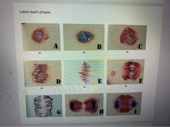
The structure of an. Ppt comparing animal and plant cell microscope lab powerpoint presentation id 842712. Cells from the Chinese Hamster Ovary are shown undergoing mitosis.
What Is A Diagram Of A Plant And Animal Cell Under An Electron Microscope Quora

Animal Cell Science Quiz

Plant Cell Diagram By Russell Kightley Media

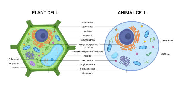
Structure Of Animal Cell And Plant Cell Under Microscope Diagrams

Magnification Bioninja

Cells Under A Microscope By Jaimarie Nelson
Plant Cell Animal Cell Stock Illustrations 760 Plant Cell Animal Cell Stock Illustrations Vectors Clipart Dreamstime
3 1 How Cells Are Studied Concepts Of Biology 1st Canadian Edition
Solved Ectron Micrographs Of Animal And Plant Cell Our Chegg Com
Mitosis Animal Cell Under Microscope Stock Photo 1133754230 Shutterstock
Cell Structure
Solved The Image Below Are Of Cells In Mitosis That You Chegg Com
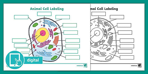
Picture Of Animal Cell Labeling Activity Digital Resources
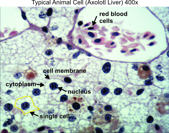
Typical Animal Cell 400x Dissection Connection
Here S How Plant And Animal Cells Are Different Howstuffworks
7 249 Plant Cell Illustrations Clip Art Istock
Lab The Cell The Biology Primer

Cells Under A Microscope By Jaimarie Nelson

Draw A Diagram Of Animal Cell And Label Any Three Parts Which Differentiate It From Plant Cell Brainly In